
Clayray Dental Radiology
57 Years
in Business
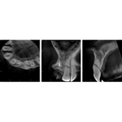
Clayray Dental Radiology runs a Morita CBCT machine that employs limited field scanning with industry leading resolution.
Clayray Dental Radiology provides detailed and advanced dental x-ray services to make life easier for dentists. Combining our technical knowledge in 3-D Cone Beam CT reconstruction with over 20 years' experience in dental, we can help make a positive difference to your treatment planning process. ? We work with dental and oral surgeons throughout Melbourne to deliver high-quality, contrast-rich Cone Beam CT imagery for dental implant planning. We have the technical and anatomical know-how to communicate our diagnostics to any dental specialist – in their language – so there’s no second-guessing and no wasting time.
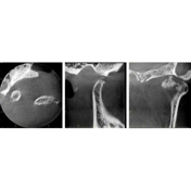
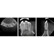
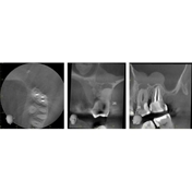
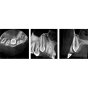
Cone Beam CT: *Implant survey to determine bony dimensions, topography, and cortical plates, *Assessment of impacted, supernumerary teeth and dens-in-dente, *Inferior alveolar canal localisation for wisdom teeth extraction, *Diagnosis of periapical infection, unfilled root canals, root fractures, *Diagnosis of periapical related pathology, *Diagnosis of temporomandibular joint disease.
What is Cone Beam CT??
Cone Beam CT is a safe and effective dental imaging technique within the dental industry, providing the clearest images available. We were the first Radiology practice to introduce CBCT technology to Melbourne & Victoria and we have become one of the most experienced with over 20 years in dental imaging. We provide patients with J-Morita CBCT scans, which is a brand of cone beam technology just like the i-Cat is.
What are the uses of Cone Beam CT?
Determining the bucco-lingual dimension, investigating the morphology of a root, localizing the inferior alveolar nerve are just some of the uses of Cone Beam examinations. In most cases they are used to aid and confirm clinical diagnosis where other imaging options have been exhausted or are inconclusive.
What is a Cone Beam CT examination?
It's an examination that produces images in 3 planes with higher resolution images and lower amounts of radiation dose when compared to medical CT scanners. Cone beam CT examinations produce geometrically accurate images that can be viewed from 3 planes. Orthopantomography and intraoral examinations in comparison only permit the viewing of a structure from 1 plane at a time with varying degrees of geometrical innaccuracy depending on how well the examination was carried out.